New neuron-holder gives life for months
 Bio-engineers have created a brain-like tissue that shares some functions with our own grey matter, and they kept it alive in a lab for over two months.
The breakthrough that made the brain-like tissue possible was the creation of a novel composite structure, which combined natural substances to encourage brain cells to grow.
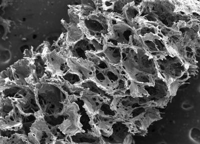
The composite consisted of two biomaterials with different physical properties: a spongy scaffold made out of silk protein and a softer, collagen-based gel.
The scaffold served as a structure onto which neurons could anchor themselves, and the gel encouraged axons to grow through it.
The researchers cut the spongy scaffold into a doughnut shape and populated it with rat neurons.
They then filled the middle of the doughnut with the collagen-based gel, which subsequently permeated the scaffold.
In just a few days, the neurons formed functional networks around the pores of the scaffold, and sent longer axon projections through the centre gel to connect with neurons on the opposite side of the doughnut.
The result was a distinct ‘white matter’ region (containing mostly cellular projections, the axons) formed in the centre of the donut that was separate from the surrounding ‘grey matter’ (where the cell bodies were concentrated).
Over the following few weeks, researchers conducted experiments to determine how well the neurons were growing in their brain-like tissue, compared with neurons grown in a collagen gel-only environment or in a 2D dish.
The researchers found that the neurons in the 3D brain-like tissues had higher expression of genes involved in neuron growth and function.
Additionally, the neurons grown in the 3D brain-like tissue maintained stable metabolic activity for up to five weeks, while the health of neurons grown in the gel-only environment began to deteriorate within 24 hours.
Neurons in the 3D brain-like tissue exhibited electrical activity and responsiveness that mimic signals seen in the intact brain, including a typical electrophysiological response pattern to a neurotoxin.
Researchers sought to determine whether they could use the tissue to study traumatic brain injury.
To simulate a traumatic brain injury, a weight was dropped onto the brain-like tissue from varying heights and recorded changes in the neurons' electrical and chemical activity.
The tissue was developed at Tufts University in Boston by a team led by Professor David Kaplan.
Kaplan says the ability to study traumatic injury in a tissue model offers advantages over animal studies, in which measurements are delayed while the brain is being dissected and prepared for experiments.
“With the system we have, you can essentially track the tissue response to traumatic brain injury in real time,” said Kaplan.
 “Most importantly, you can also start to track repair and what happens over longer periods of time.”
Kaplan says the brain-like tissue's longevity will be hugely important for studying other disorders.
“The fact that we can maintain this tissue for months in the lab means we can start to look at neurological diseases in ways that you can't otherwise because you need long timeframes to study some of the key brain diseases,” he said.


 Print
Print